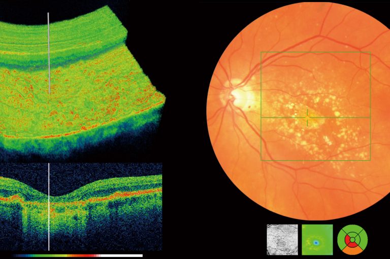
OCT
Ocular ultrasonography
- Biometry (color-coded-3 dimension)
- Flourescien angiography
- Indocyanine green angiography
- Electrophysiological green angiography
- Electrophysiological tests VEP, ERG, EOG
- Automated preimetry
- Corneal Topography
- Corneal pachymetry
- OCT
- Anterior Segment OCT
Perimetry is the measurement of he visual functions of the eye at topographically defined loci in the visual field. the visual field is hat portion of the external environment of the observer wherein the steadily fixing eyes can detect visual stimuli.
We use 101 automated static perimery which proved to be a sensitive test in many ophtalmological and neurological conditions.
There are several reasons for doing the first optic disc as cupping, pallor, edoema or asymmetry, APD, history or family history of glaucoma, diabetes, transient visual loss, neurological conditions as MS and brain tumors, or infractions or high myopia or headache.
And once field defects have been documents, it is necessary that the patient needs to be reexamined regularly every 3-12 months according to the condition of the patient.
Special Investigations:
- Fluorecein Angiography
- Fundus Green Angiography
- Indocyanine Green Angiography
- Ultrasonography
- Ultrasonography
- Biomery
- Preimetry